


Introduction
Tissue engineering, as one of the most promising and challenging frontiers in medical life sciences, has attracted extensive attention from the academic community due to its unique advantages in repairing injuries and restoring functions, as well as its significant social and economic value. The core components of this field include seed cells, biomaterials, and tissue construction. Among them, tissue engineering scaffolds—materials capable of integrating with living cells and being implanted into biological systems—serve as three-dimensional supports for seed cells before they form functional tissues, providing essential spatial environments for cellular proliferation, differentiation, nutrient exchange, metabolism, and extracellular matrix secretion.
In recent years, polylactic acid (PLA) and poly(lactic-co-glycolic) acid (PLGA) have emerged as key research directions in the field of tissue engineering biomaterials due to their excellent biocompatibility, biodegradability, and dual functionality as both structural scaffolds and controlled-release carriers [1]. This article aims to provide a comprehensive review of the applications of PLGA copolymers in tissue engineering research.
Structure and Degradation Mechanism of PLGA Copolymers
PLGA is synthesized by block copolymerization of lactide (the cyclic dimer of lactic acid, LA) and glycolide (the cyclic dimer of glycolic acid, GA) in varying ratios. During synthesis, the carboxyl (-COOH) and hydroxyl (-OH) groups of LA and GA monomers undergo esterification reactions, forming linear chains connected via ester bonds (-COO-). Key molecular characteristics of PLGA—including molecular weight and distribution, LA/GA ratio, sequence distribution pattern, end-capping functional groups, glass transition temperature, and morphology—directly influence its material properties [2].
Illustration: Schematic representation of the synthesis of poly(lactic-co-glycolic) acid (PLGA)
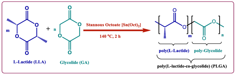
The molecular weight of PLGA can be adjusted based on the ratio and molar mass of LA and GA monomers. When the molecular weight is below 5000 Da, PLGA appears viscous at room temperature, becoming increasingly viscous as the molecular weight decreases. PLGA with molecular weights between 5000 and 10,000 Da tends to form solid blocks at room temperature. PLGA with molecular weights ranging from 10,000 to 15,000 Da typically exists as a powder, while PLGA exceeding 15,000 Da generally forms irregular particles. Generally, PLGA with a molecular weight above 30,000 Da is suitable for long-term implantable devices, whereas medium-molecular-weight PLGA (e.g., 5000–30,000 Da) is more appropriate for fabricating porous cell scaffolds or sutures.
The degradation of PLGA occurs through hydrolysis of the ester bonds in its main chain, producing lactic acid and glycolic acid as degradation products. In vivo, PLGA first breaks down into monomeric lactic acid, which enters the tricarboxylic acid cycle, while glycolic acid is either excreted via the kidneys or metabolized through the same cycle. The degradation rate of PLGA copolymers is influenced by multiple internal and external factors:
(1) Molecular weight and its distribution. A broader molecular weight distribution results in a higher concentration of terminal carboxyl groups, promoting autocatalytic degradation; conversely, narrow molecular weight distributions contain fewer terminal carboxyl groups, leading to slower degradation rates.
(2) LA/GA ratio. The properties of PLGA vary significantly depending on the molar ratio of LA to GA. Studies show that higher LA content leads to slower degradation rates. For example, when the LA:GA ratio is 10:90, degradation may take 3–6 months; increasing the LA ratio to over 20% extends the degradation time to 12–24 months, while simultaneously enhancing mechanical strength and reducing flexibility. Conversely, higher GA content results in faster degradation and greater flexibility [3].
(3) The effect of acidity and alkalinity on the degradation rate of PLGA. Some researchers have found that during in vitro degradation, PLGA copolymers produce lactic acid, leading to a local decrease in pH and triggering an autocatalytic phenomenon that accelerates degradation [4]. Maulding et al. [5] reported that acidic tertiary amine compounds such as methyldopa contained within the copolymer can accelerate microsphere degradation, whereas basic compounds can bind with carboxyl groups, thereby reducing the degradation rate. Therefore, when designing and preparing PLGA copolymers, the influence of acidic or basic compounds should be taken into account.
(4) The effect of PLGA material shape on its degradation. Pistner et al. [6] reported implanting two types of implants made from the same batch of PLGA material subcutaneously in mice: one was a semi-crystalline open structure, and the other was an amorphous, dense structure. Results showed that the degradation behavior of shaped materials differed from that of amorphous and crystalline materials. In amorphous materials, the semi-crystalline nature of the copolymer allows low-molecular-weight components to degrade through the amorphous phase, while shaped materials do not allow this pathway and instead degrade primarily via autocatalysis. For semi-crystalline polyesters, degradation initially occurs in the amorphous regions, followed by the crystalline regions.
Requirements for PLGA Copolymers in Tissue Engineering
In tissue engineering, ideal carrier materials should meet the following criteria:
(1) Be fully degradable, with a degradation rate matching the regeneration speed of seeded cells;
(2) Exhibit excellent biocompatibility, be non-toxic and non-teratogenic, and promote cell adhesion and proliferation;
(3) Have a porosity greater than 90%, pore size between 100–500 μm, and possess a three-dimensional porous structure with interconnected pores. This enables new tissue ingrowth and gradual degradation of the material, facilitating vascularization and providing sufficient mechanical strength;
(4) Maintain the structural integrity and phenotype of seeded cells, offer effective surface activity, provide an optimal microenvironment for cell growth, proliferation, and extracellular matrix secretion, activate cell-specific gene expression, sustain normal cellular phenotypes, and induce tissue formation;
(5) Biomaterial research must be closely integrated with tissue construction, which is the ultimate goal of tissue engineering biomaterials. Many challenges encountered during tissue construction depend on advances in biomaterial science;
(6) The chemical properties and processing techniques of the material determine its functional characteristics. Thus, biomaterials should possess adequate moldability for easy shaping, sufficient mechanical strength to maintain their form over a certain period after implantation, ensuring the resulting tissue achieves the desired shape [7–8].
PLGA copolymers have become excellent polymer-based, biodegradable carrier materials in tissue engineering precisely because they fulfill these fundamental requirements.
Applications of PLGA Copolymers
In recent years, the rapid development of biodegradable biomaterials has led to widespread medical applications of polylactic acid-based polymers, including surgical sutures [9], drug-controlled release systems [10], orthopedic fixation [11], and tissue repair materials [12]. These have become among the most widely used biomaterials for cell scaffolds and have been included in the U.S. Food and Drug Administration (FDA) pharmacopoeia [13].
PLGA copolymers were first applied in orthopedics and dentistry. Later, it was discovered that PLGA exhibits excellent tissue compatibility, protects biological proteins from enzymatic degradation, and provides controlled drug release, maintaining effective local drug concentrations over extended periods. As a result, PLGA has recently become a key focus in pharmaceutical research on sustained-release drug delivery systems [14].
PLGA copolymers can also serve as scaffolds for tissue cells, inducing the production or release of tissue factors, and are therefore widely used in oncology, trauma repair and aesthetic medicine, neurology, dermatology, and various other clinical disciplines. Stoll et al. [15] investigated the role of PLGA's three-dimensional scaffold structure in treating tendon rupture healing. The results showed that the 3D PLGA scaffold provided an excellent growth environment for tendon cells and extracellular matrix, promoting tendon cell proliferation and regeneration, reducing tendon adhesions, and facilitating tendon healing.
Applications of PLGA Copolymers in Tissue Engineering
As a promising biodegradable polymer material, PLGA has been extensively applied as a scaffold in tissue engineering.
Kang et al. [16] injected porous and non-porous PLGA microspheres containing rabbit chondrocytes subcutaneously into nude mice. After six weeks, both groups formed white, firm cartilage tissues. Histological analysis revealed that the cross-sectional area of the porous PLGA group was significantly larger than that of the non-porous group, and the content of glycosaminoglycans and collagen in the porous PLGA group was markedly higher. The study demonstrated that open-pore PLGA microspheres favor cartilage regeneration and are suitable for constructing cartilage tissue in tissue engineering.
Bone morphogenetic proteins (BMPs) can irreversibly induce undifferentiated mesenchymal stem cells to differentiate into chondrocytes and osteoblasts, making them key growth factors for bone defect repair and reconstruction. However, when administered alone, BMPs are rapidly lost, degraded, and absorbed in vivo, limiting their ability to effectively exert bone-inductive effects. Therefore, synthetic PLA/PLGA biomaterials are commonly used as scaffolds to deliver BMPs in bone regeneration research.
Kenley et al. [17] conducted skull defect repair in mice by implanting BMP/PLGA-MS into 8 mm diameter cranial defects. After 21 days, new bone formation was observed at both the inner and outer cortical layers, along with the development of marrow tissue, and the newly formed bone maintained the normal contour of the skull.
Li et al. [18] developed a highly porous scaffold composed of ultrafine fibers made from polylactic-glycolic acid copolymer (PLGAC), with diameters ranging from 500 to 800 nm. The scaffold exhibited a porosity exceeding 90%, good mechanical properties, and a morphology resembling the extracellular matrix of living tissues. In addition to significantly enhancing cell adhesion and proliferation, it effectively preserved cell morphology and guided directional cell growth along the fiber orientation. This cellular scaffold closely resembles the extracellular matrix (ECM) in structure, demonstrates excellent biocompatibility, meets design requirements for cell scaffolds, and represents a promising biomaterial choice in tissue engineering.
Wang Gongxian et al. [19] studied the effect of MSCs combined with PLGA scaffolds on repairing damaged skin tissue. Cell-scaffold complexes (incubated for 12 hours) and PLGA scaffolds were transplanted onto rabbit skin wounds. Four weeks post-surgery, gross observation revealed significant wound contraction and sparse hair growth on the newly formed skin in the group treated with PLGA alone; in contrast, the MSC-PLGA composite group showed minimal wound contraction and a surface appearance nearly identical to normal skin.
Histological examination showed that the newly formed skin in the MSC-PLGA group had a structure similar to normal skin, including well-developed epidermal, subcutaneous, and dermal layers, and contained functional skin appendages such as hair follicles, sebaceous glands, and sweat glands. Newly formed skin derived from PLGA scaffolds alone also showed the development of epidermal, subcutaneous, and dermal layers. However, thickened fibrous scarring was observed within the dermis, and apart from a few hair follicles, no other appendages were formed.
These studies highlight the promising clinical potential of PLGA-based tissue engineering with seeded cells for regeneration of bone, cartilage, and skin. Nevertheless, challenges such as uneven degradation, mismatch between degradation rate and tissue formation rate, inflammation caused by acidic byproducts from localized degradation, poor water absorption, and weak cell adhesion capacity have hindered further application of PLGA in tissue engineering research, necessitating more in-depth investigation.
References
[1] Liu H, Slamovich EB, Webster TJ. Less harmful acidic degradation of poly(lactic-co-glycolic acid) bone tissue engineering scaffolds through titania nanoparticle addition[J]. Int J Nanomedicine, 2006, 1(4):541-545.
[2]EL-HAMMADI MM, ARIAS JL. Recent advances in the surface functionalization of PLGA-based nanomedicines[J]. Nanomateri, 2022, 12(3): 354.
[3] TAŞKOR ÖNEL G. Synthesis and characterization of poly(lactic-co-glycolic acid) derived with L-glutamic acid and L-aspartic acid[J]. Erzincan Univ J Sci Technol, 2023, 16(1): 155.
[4] Alpaslan C, Irie K, Takahashi K, et al. Long-term evaluation of recombinant human bone morphogenetic protein-2 induced bone formation with a biological and synthetic delivery system[J]. Br J Oral Maxillofac Surg, 1996, 34(5): 414–418.
[5] Maulding HV, Tice TR, Cowsar DR, et al. Biodegradable microcapsules: acceleration of polymeric excipient hydrolytic rate by incorporation of a basic medicament[J]. J Contr Rel, 1986, 3(1–4): 103–117.
[6] Pistner H, Bendix DR, Muhing J, et al. Poly(L-lactide): a long-term degradation study in vivo. Part III. Analytical characterization[J]. Biomaterials, 1993, 14(4): 291–304.
[7] Zhang Jianxin, Xu Zhanwang, Chang Feng. Experimental study on tissue-engineered artificial bone for bone defect repair[J]. Chinese Journal of Orthopedics, 2009, 17(16): 1258–1261.
[8]Hollister SJ.Porous scaffold